Macroscopic images example
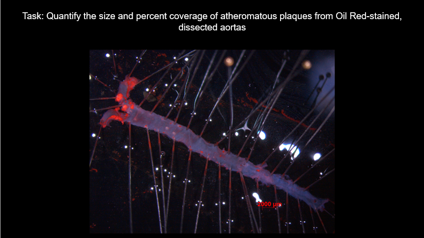
Example showing recognition of aorta, identification and area quantification of stained atheromatous plaques (using a plugin written in ImageJ Macro Language)
Flurescence microscopy example
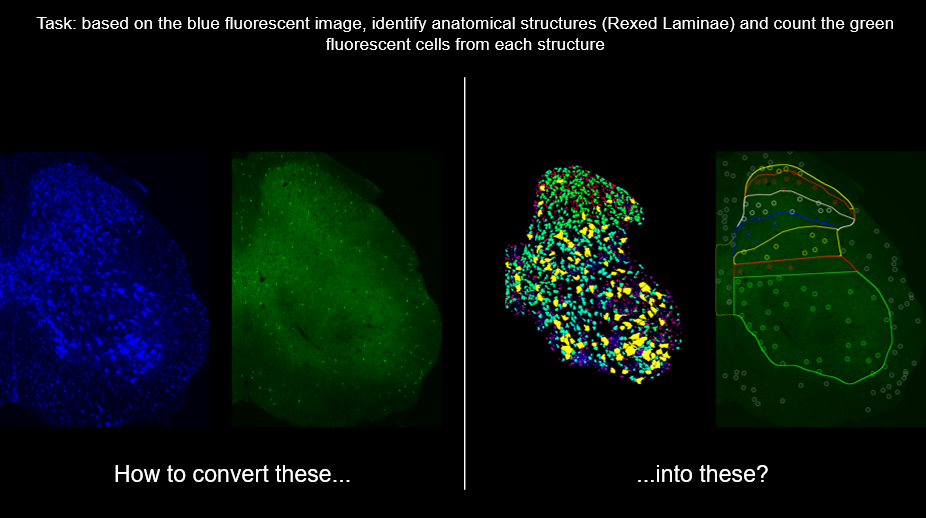
Example showing anatomical structures’ identification from micrographs and usage to count specific cells in the regions
Other examples of image analysis
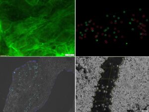
More examples for automated image analysis: scratch assay, fiber orientation, percent positive cells, surface/interior cells